
Neurons
There is also a screencast of a version of this lecture using the book's slide deck found here.
Here is a slide presentation from Campbell on the topic.
First, the overall system:
We break the system down by a couple of overlapping categories. First we have the Central Nervous System (brain and spinal cord) and the Peripheral Nervous System (connections out to the body). A “nerve” will contain a bundle of nerve cells known as “neurons.” So, a nerve is sort of a conduit with many wires.
The neurons in the PNS may carry signals from the body back to the CNS (sensory or afferent neurons), or from the CNS to the PNS (motor or efferent neurons). There are also “interneurons,” which serve as connectors, primarily in the CNS.
What you control and what you don’t
The PNS will have a set of neurons of whose signals you are aware and/or you control. This voluntary muscle control system is called the somatic nervous system (SoNS). In the CNS, there are also parts of the brain that are part of your voluntary system.
There are also parts of the brain and spinal cord that are not under your control and of which you are not conscious. These connect with the autonomic nervous system in the peripheral system. These neurons sense and control systems such as your pupil dilation, heart rate and breathing, digestive system and more than you really know about including control of hormones.
The autonomic system is broken down further into the Sympathetic and Parasympathetic systems. These systems often work toward opposite goals, not so much antagonistically, but in a complementary manner. Their interaction is critical in homeostasis.
They also differ in the subsets of neurotransmitters and receptors they use.
Sympathetic:
Often thought of as controlling the “Fight or Flight” response. Signals from this system result in things you associate with when you feel threatened. Your pulse increases, as does blood pressure. Flow of blood and nutrients to the extremities increases at the expense of internal organs (including most of the brain). You are unlikely to notice pain as much. Any other urges (hunger, feeling tired, sex drive) all take a back seat to getting out of trouble.
The neurotransmitters most associated with this system are norepinephrine, epinephrine (also known as noradrenalin and adrenaline) and acetylcholine. Epinephrine and norepinephrine are also hormones…more on that later. The acetylcholine receptors in this system tend to be of a class that also binds the nicotinic acid molecule.
All of these signals are associated with the fight or flight response.
Parasympathetic:
These are your “rest and digest” and “feed and breed” responses (if prefer: "Netflix and Chill). If hunger, sex drive, tiredness etc go out the window when you are running for your life, these are the signals that counteract this response.
The neurotransmitters also include acetylcholine (but usually a different receptor) and GABA (gamma amino butyric acid).
Neurons
Below is the labeled diagram from the board.
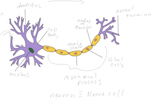
It has most of the terms we discussed. The direction of impulse travel is from left to right in the neuron given. While this is a “typical” rendition, Other shapes occur.
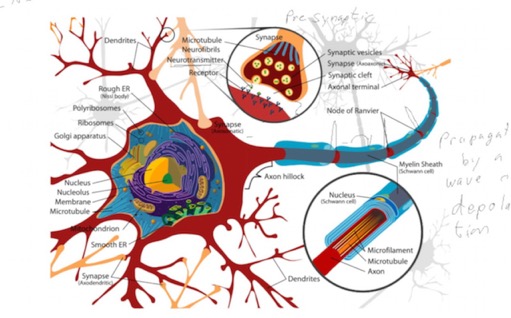
This shows some of the detail of the cytoskeleton, which is rather specialized. Most of the other components you recognized from a typical cell (whatever that means). Note the myelin sheath, made from glial cells, which wraps the axon at regular intervals (not all neurons are myelinated). The nodes between the myelin cells are really what’s important in transmitting an action potential down the axon.
The Synapse
There is nothing really new here: The "pre-synaptic" cell is justing doing stimulated secretion, which works via calcium-stimulate fusion of vesicles at the plasma membrane (mediated by "snap 25," which we met early in the year). The post-synaptic cells is just doing chemical reception.
There are some new things that allow for the regulation of these signals…such as ligand "reuptake" receptors. You may have heard of drugs such as SSRIs, Selective Serotonin reuptake inhibitors.
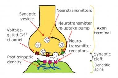
Here is a closer view of the synapse. The two cells are held in close apposition to each other by proteins not shown. The presynaptic cell, the one sending the signal, releases a neurotransmitter (not conceptually different from any signal molecule). It binds to a receptor on the receiving cell (post synaptic cell), and initiates a response. Again, not very different from any signal event. The response is to initiate an action potential, by activating a channel. There are feedback pathways to make sure the signals stay at the right level. Also, there are specific pathways to remove the neurotransmitter from the cleft between the cells. This is done either by a “re-uptake” receptor, which does what the name implies, or by degrading it enzymatically. The voltage-gated calcium channels shown in the presynaptic cells is one way to initiate the release of neurotransmitters.
This is a link to an amazing reconstruction of a synapse.
Action Potential
We’re going to do a simple version of this really. The time scale of the reaction is milliseconds or less.
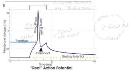
It starts with an electrochemical gradient, which all cells have. It’s larger in neurons such that the voltage across the membrane is about -70 millivolts. The gradient is established by pumps like the ones we discussed earlier in the year that pump sodium, potassium calcium and chloride ions asymmetrically across the membrane. There is more Na+ outside the cell than inside. More potassium outside. Since it carries a positive charge, the inside of the cell is net negative in charge. When channels are opened, ions will flow down their electrochemical gradient (attempting to equalize both charge and chemical distribution).
The two main players are ligand-gated sodium channels and voltage-gated sodium channels.
The ligand-gated channels open in response to neurotransmitter. The more neurotransmitter released, the more the ligand-gated channels open. That allows Na+ to flow down gradient and pushes the voltage toward neutral. If it is too weak of a signal, the potential never gets above the threshold shown, and the neuron doesn’t “fire.” However, if enough of the ligand-gated channels open, the voltage reaches threshold and that change in voltage opens the voltage-gated channels.
Voltage-gated channels.
These are special protein structures. They have three states:
- closed and ready to respond to threshold voltage
- open, allowing ions through
- “refractory,” which is closed and not able to respond to voltage
I’m going to link to a good website that shows you the sequence of events.
Have to get to some other things.